For many years, the Plant Biotechnology and Micropropagation Team at the Botanical Garden has been engaged in the study of somatic embryogenesis, which is one of the most effective routes of in vitro plant reproduction. In this process, progeny organisms arise directly from the cells of the plant's body - without a fertilization phase, a phenomenon peculiar to the plant world. One of the key factors determining the success of regeneration by this route is the selection of the initial tissue (the so-called explant).
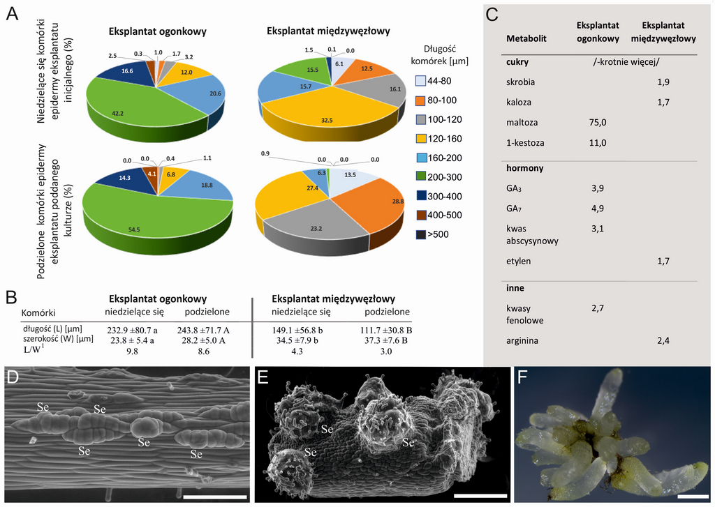
In the present study, we compared two types of tree fern explants Cyathea delgadii taken from the petiole and interveinal section, yielding somatic embryos of unicellular and multicellular origin, respectively. Using a variety of microscopic techniques, we examined the morphology and structure of the initial explants and the cells that build them. Meanwhile, using high-performance liquid chromatography, we determined the levels of 89 endogenous metabolites, i.e. sugars, hormones, phenolic acids, polyamines and amino acids.
We showed that the high content of maltose, 1-kestose, abscisic acid, biologically active gibberellins (GA3 i GA7) and phenolic acids were characteristic of the formation of somatic embryos via the single-cell pathway. In turn, high levels of starch, calose, ethylene and arginine favored their multicellular origin. The work presented here is the first such detailed presentation of the characteristics of the initial explant (Figure 1), which may play an important role in the induction of somatic embryogenesis and condition the pathway of somatic embryo formation.

Figure 1. Comparison of petiole and internode explants. (A) Percentage of cells in relation to their length. (B) Average cell length and width: non-dividing epidermal cells of initial explants; divided epidermal cells of explants during in vitro culture. (C) Differences in the content of selected metabolites that may indicate the formation of somatic embryos by a single-cell (caudal explant) or multicellular (interstitial explant) route. (D, E) Somatic embryos developed from single cells of the petiole explant epidermis (D) and from multiple adjacent cells of the internode explant (E); images obtained from a scanning electron microscope. (F) Juvenile sporophytes obtained by somatic embryogenesis from single cells of the petiole explant epidermis after 2 months of culture conducted in the dark; images obtained from a stereo light microscope. Abbreviations: GA3, GA7 - Gibberellins; Se - somatic embryo. Scale: (D) 200 µm; (E) 500 µm; (F) 1 mm. 1 The L/W ratio was calculated as the average ratio of length and width for each cell.
Mikula A, Tomaszewicz W, Dziurka M, Kazmierczak A, Mushroom M, Sobczak M, Zdańkowski P, Rybczynski J. 2021. the Origin of the Cyathea delgadii Sternb. Somatic Embryos Is Determined by the Developmental State of Donor Tissue and Mutual Balance of Selected Metabolites. Cells 10(6), 1388; https://doi.org/10.3390/cells10061388
post
post
2021-06-23 05:12:39